AI Image Detector for Research Figures and Scientific Papers
 Sana Bano
·October 8, 2026
·17 min read
Sana Bano
·October 8, 2026
·17 min read
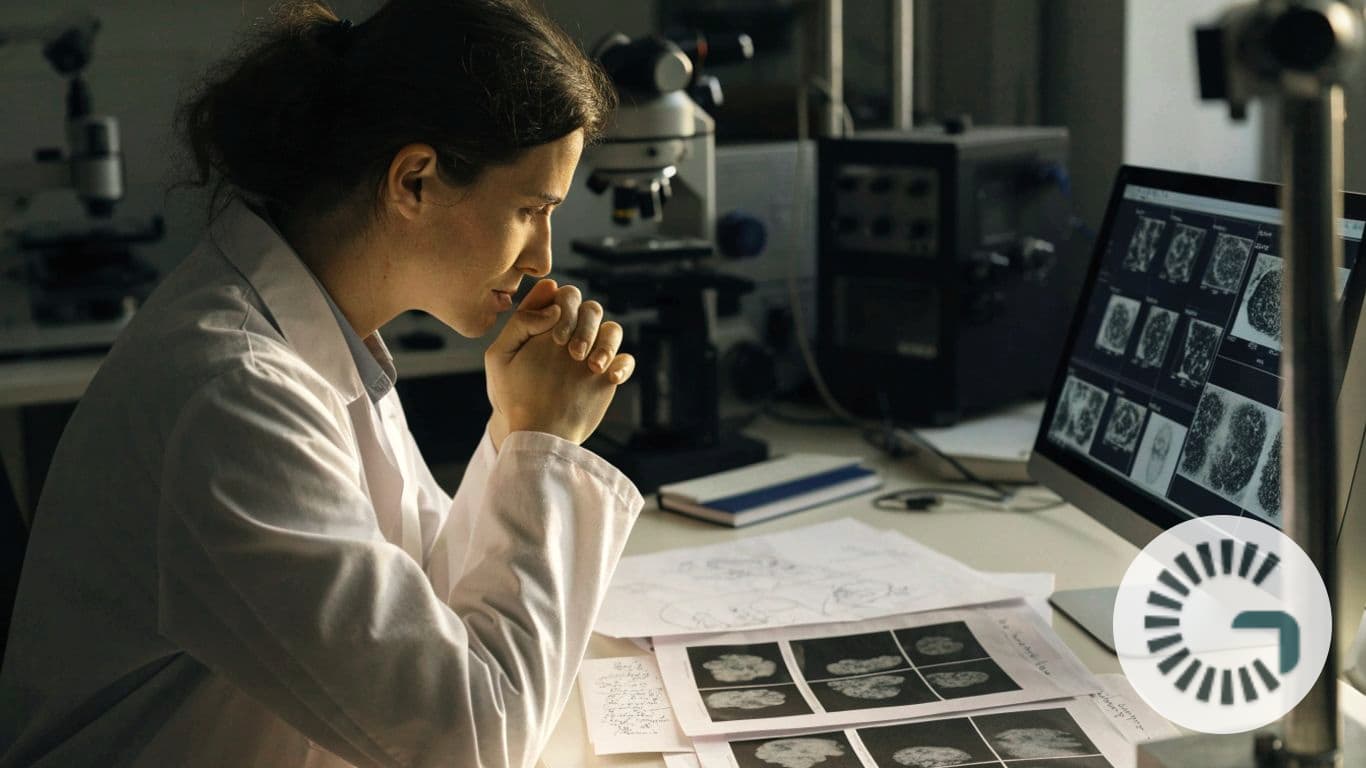
Use an ai image detector for research figures and scientific papers as a screening signal, not proof.
Key Takeaways
An AI detector can help you decide which research images deserve a closer look, but it cannot determine misconduct on its own. Use its results alongside original files, experiment records, and careful human review.
- Detection can flag signs of AI generation or image alteration, but these are different questions from whether a figure is scientifically valid.
- A score or highlighted region is a reason to investigate, not proof of how an image was made.
- Scientific image types, file quality, and routine processing can all affect detector results.
- Original images, processing notes, and consistent review steps make follow-up more useful.
- Any decision about a research concern should follow the relevant journal or institutional policy.
What an AI image detector for research figures and scientific papers can identify
An ai image detector for research figures and scientific papers can help you screen visuals for signs that merit a closer review. It may assess whether an image appears AI-generated or altered, but those are distinct questions, and neither result alone establishes misconduct. The right interpretation depends on the image’s role, the study context, and what original material is available. Treat screening as a starting point for inquiry, not a verdict.
AI-generated images versus altered or duplicated figures
AI generation, editing, and duplication are related but separate concerns. A generated image may be synthetic from the outset, while an altered image may begin with genuine data and later be modified; a duplicated panel may reuse all or part of another image. GPTOne AI Image Detector assesses whether images appear real, AI-generated, or AI-modified. That scope can inform an authenticity check, but it does not by itself determine whether a particular reuse or edit was appropriate in its scientific context.
Common image types in scientific papers, from microscopy to medical imaging
Research papers contain many kinds of visuals: microscopy fields, gel images, scans, photographs, charts, and assembled multi-panel figures. They differ in texture, acquisition, resolution, and how much processing is customary, so a detector that performs well on one kind of image may be less informative on another. Before relying on a result, check whether the tool has been assessed on the types of figures you actually review. A visually unusual pattern in a scan, for example, needs different contextual review than a similar pattern in a photograph.
What detection results can and cannot establish
A detector result can point to signals in an image that deserve attention. It cannot establish who created the image, whether a particular transformation was disclosed, or whether the underlying experiment was conducted as reported. Confidence estimates should be read as model outputs, not as calibrated probabilities of misconduct unless validation supports that specific interpretation. For a practical overview of detector signals and limits, focus on what the result shows and what it leaves unanswered.
Why image screening is only one part of research integrity review
A figure is part of a larger evidentiary record. You may need to compare it with raw files, laboratory notes, figure legends, methods, and earlier versions of a manuscript before you can understand a concern. A detector can help direct attention, but scientific judgment and procedural fairness still matter. The same careful approach applies to automated systems generally; see this caution about detector limits when deciding how much weight to give a score.
How AI image detection works
Image detectors look for patterns that may differ between naturally captured, edited, and generated images. Methods vary, and a result can reflect several signals rather than one decisive clue. Some tools also examine file information or provenance records, when present. Understanding the kinds of evidence a detector uses helps you ask better questions about an alert.

Visual patterns and artifacts detectors may analyze
A model may assess visual and frequency-based patterns, including texture, noise, or inconsistencies in local regions. These features can be subtle and may be difficult to judge by eye, especially after figures have been resized or assembled. GPTOne AI Image Detector combines model-based detection with visual and frequency-based signals, metadata and generator fingerprints, and provenance checks such as C2PA Content Credentials. Its output is still an assessment of signals, not a confirmation of what happened during an experiment. For more context, you can try the image detector and treat any result as one part of a review.
Metadata and provenance signals that can support an assessment
Metadata can sometimes preserve useful details about a file, such as its format or information recorded by software. Provenance credentials, when present and valid, may add context about an image’s history. But these records can be absent, stripped during export, or incomplete, so their absence is not evidence of manipulation. A responsible review distinguishes between a missing record and a positive signal, then checks whether the original file or another reliable source can answer the question.
Why different models can produce different results
Detection systems may be trained or configured differently, and they may respond to different image characteristics. As a result, two tools can give different assessments of the same figure without either result settling the matter. Training examples, supported image types, thresholds, and the way a score is presented all affect what you see. Where results conflict, record the disagreement and return to the source material rather than selecting whichever output confirms an initial suspicion.
How image transformations can affect detection
A figure often passes through several ordinary steps before publication: cropping, resizing, contrast adjustment, conversion, or layout in a composite panel. Each can change pixel patterns or remove information that a detector might otherwise use. Keep the submitted image and, where possible, earlier source files so you can compare versions. If a flag appears only after export or compression, that detail can help explain the result, though it does not automatically resolve every concern.
Where detectors can help in the research workflow
Screening is most useful when it fits into a clear review process rather than being applied as an isolated test. Authors, editors, and research teams may use it at different stages, with different responsibilities and different access to source records. The aim is to identify questions early and route them to the people equipped to assess them. A consistent process also helps prevent a score from being treated differently depending on who submitted the work.
Screening figures before manuscript submission
Before submission, you can use screening as a final check that prompts you to review the figures and supporting files. If an image is flagged, confirm that the figure matches the underlying data and that any permitted processing is documented. This is a chance to catch missing source files or unclear figure assembly notes while you can still resolve them. It is not a reason to alter an image simply to change a detector’s result.
Reviewing image concerns during peer review
During peer review, a flag should be handled with care because reviewers may not have access to raw data or the full history of a figure. Describe the specific visual concern, avoid claims about intent, and follow the journal’s process for confidential communication. A detector can help narrow attention to a region, but it should not replace expert assessment of the scientific material. Keep the language factual: identify what appears unusual and what evidence would help clarify it.
Checking published work during research integrity investigations
When a concern arises after publication, the image should be evaluated alongside the paper and its supporting record. A detector result may help prioritize which figure or region to examine, but investigators need access to relevant source material and a fair process for seeking an explanation. Preserve the image as reviewed and note any transformations made for analysis. Conclusions belong to the appropriate review process, not to an automated score alone.
Applying screening consistently across a lab or journal
Consistency matters because different thresholds or ad hoc decisions can lead to uneven treatment. A written workflow can define when screening happens, who reviews a flag, and what evidence is needed before escalation. For example, a journal might distinguish a routine technical query from a potential integrity concern and route each appropriately. Clear roles make screening more useful without turning it into a blanket accusation.
How to evaluate an AI image detector for scientific use
The best detector for your setting is not necessarily the one with the most confident-looking result. You need to know what image types it covers, how it was evaluated, what happens to submitted files, and whether a human can inspect the basis for a flag. Those practical details matter especially when unpublished work or sensitive research is involved. A careful evaluation begins with your actual workflow, not a general claim of accuracy.
Coverage across image modalities and file formats
Check whether a tool accepts the formats and image types your team encounters, and whether its stated scope matches the figures you plan to screen. A general image detector should not be assumed to work equally well on every modality or on complex multi-panel figures. This simple comparison can help you ask focused questions before adopting any tool.
| Review question | Why it matters |
|---|---|
| Which image types are included? | Performance may vary across scientific modalities. |
| Which file formats are accepted? | Conversion can change or remove image information. |
| Can it assess a full figure or individual panels? | Your workflow may require one or both. |
| What are the stated exclusions? | Unsupported cases should not be treated as cleared. |
Use the answers to define where a tool fits and where manual review remains necessary. If the vendor’s documentation does not address an image type you routinely handle, consider that an unknown rather than an implicit assurance.
Validation data, benchmarks, and known limitations
Ask how the tool was tested, what examples were included, and whether the evaluation resembles the images in your own collection. A benchmark on ordinary photographs may say little about research figures with overlays, stains, scans, or multiple assembled panels. Look for clear discussion of false positives, missed detections, and changes in performance after image processing. When a provider does not explain its validation, you should be cautious about treating a score as comparable across tools or use cases.
Privacy, data retention, and manuscript confidentiality
Before uploading unpublished figures, review where analysis takes place, how files are handled, and whether they are retained. GPTOne AI Image Detector is designed to run image analysis locally in the browser, supporting a privacy-focused workflow. Still, you should confirm that the tool and your institution’s rules fit the sensitivity of the material you plan to review. Privacy expectations are not a substitute for checking applicable policies or contractual terms.
Reporting features and options for human review
A useful report should help you inspect a concern rather than obscure it behind a single number. Look for a clear confidence score or signal breakdown, and for a way to see which regions may warrant attention if the tool offers one. GPTOne’s image analysis can include an AI-vs-real confidence score, detailed signal analysis, and a region heatmap highlighting potentially manipulated areas. Such outputs can guide follow-up, but you still need the original image and a qualified reviewer to assess what the signal means.
How to interpret detector results responsibly
A detector’s output can be compelling, especially when it uses precise percentages or colored overlays. Precision in presentation does not guarantee certainty in the underlying conclusion. Read the result as a prompt to gather context and compare evidence, not as a standalone finding. That distinction protects both the quality of the review and the people whose work is being examined.
Treating scores as signals rather than proof
A confidence score expresses the tool’s assessment under its own method; it is not automatically the probability that an image was generated, altered, or used improperly. The score may also depend on the image quality and transformations applied before analysis. Treat it as one signal among several, and be explicit about uncertainty when communicating it. High-stakes judgments should rely on a fuller review than a single automated output.
Comparing flagged regions with original files and experiment records
If a region is highlighted, compare it with the original, higher-quality files and the records describing how the figure was prepared. Ask whether the flagged area corresponds to a known crop, an adjustment, or a panel boundary. Examine the surrounding evidence too: a localized alert may be meaningful, but it may also reflect a routine image feature. Keep the comparison focused on observable differences rather than assumptions about motive.
Checking for benign causes such as compression or image processing
Many legitimate steps can change image appearance. Before escalating a flag, identify whether export, compression, format conversion, or documented image processing could have affected it. Then check whether the same pattern exists in the original file or appears only in a later version. This context can help separate a technical artifact from an unresolved question, though it may not answer every case.
Documenting the review and its reasoning
A short record makes a review easier to understand later. Note which file was screened, which version of the tool was used if known, what it flagged, and what comparisons or follow-up took place. Include explanations that support the conclusion as well as evidence that remains uncertain. This keeps the review transparent and avoids turning a detector’s first impression into an undocumented decision.
Common sources of false positives and missed detections
No detector sees an image in a vacuum. Routine scientific processing, poor source quality, or new generation methods can all complicate interpretation. A false positive may direct attention to a legitimate feature, while a missed detection can leave a concern undiscovered. Knowing these limits helps you decide what a result can reasonably support.
Legitimate adjustments that can resemble image manipulation
Scientific images may be adjusted for presentation or assembled under rules that vary by field and publication. A contrast change applied uniformly, for example, is different from selectively changing a result, but a detector may not know the purpose or documentation behind an adjustment. Check the applicable figure policy and the author’s processing records before reaching a conclusion. The question is not simply whether pixels changed, but whether the change was permitted, transparent, and scientifically appropriate.
Cropping, resizing, compression, and figure assembly
Cropping can remove surrounding context, resizing can introduce interpolation, and compression can create visible artifacts. Combining several panels into a single figure may also produce boundaries or repeated visual patterns that are easy to misread without the source files. These operations can affect both what a detector sees and what a reviewer can verify. Compare the assembled figure with component images whenever they are available.
Low-quality scans and complex scientific imagery
Low resolution, noisy scans, staining variation, and intricate structures can make image interpretation difficult even for a knowledgeable reviewer. A detector trained on a different visual domain may respond to unusual texture without recognizing its scientific source. If the image is poor quality, seek a better copy or original rather than treating a weak signal as decisive. When the modality is specialized, involve someone familiar with how those images are produced and processed.
New generation methods that may evade existing detectors
Image-generation methods change, and detection systems may not recognize every method or later variation. A lack of a flag therefore cannot certify that an image is authentic. Keep the review anchored in provenance, source records, and scientific context, especially when a figure raises concerns for reasons beyond the detector output. Tools can support vigilance, but they cannot eliminate uncertainty.
Building a practical image-screening process
A sound screening process is clear about purpose, limits, and next steps. It should help you identify concerns without making automated flags a proxy for misconduct. Decide in advance what evidence will be preserved, who will review uncertain cases, and which policies govern decisions. These basics make the process fairer and more repeatable.
Setting clear rules for when and how to screen
Decide which figures will be screened, at what stage, and what kinds of results trigger a closer look. Apply the same rules to comparable submissions rather than screening selectively based on an author or a hunch. Explain that a flag leads to review, not an automatic rejection or allegation. Clear expectations reduce surprises and make it easier to use results consistently.
Keeping original images and processing records
The most useful detector result is easier to assess when you can compare it with the source. Establish a simple recordkeeping practice that keeps files linked to the figure version and preserves information about processing. At minimum, a research team can retain:
- Original image files in their received or captured form.
- Working copies used to prepare publication figures.
- Notes describing adjustments, crops, and panel assembly.
- The final submitted figure and relevant review correspondence.
These records help reviewers trace a visual change without relying on memory. They also make it easier to distinguish a documented workflow from an unexplained difference.
Escalating uncertain findings to qualified reviewers
If a concern remains after the first check, send it to someone qualified to assess the image type and the research context. Share the specific flag and the source material, while avoiding language that presumes intent. Depending on the setting, that may mean a senior researcher, journal editor, imaging specialist, or institutional office. Escalation should be proportionate to the evidence, and the person reviewing it should have enough context to evaluate the issue fairly.
Aligning decisions with journal and institutional policies
Policies define what processing is allowed, what must be disclosed, and how concerns should be handled. Check the rules that apply to the particular journal, institution, and research field before deciding what a result means. If the policy is unclear, seek guidance through the proper channel rather than creating a new standard from a detector’s output. A consistent process protects the integrity of the review as well as the scientific record.
Try a Careful Image Check
If you want a first-pass signal for an image, explore GPTOne AI Image Detector and use its result as a prompt for careful verification—not as a final judgment.
Conclusion
An AI image detector can make image screening more focused, but research integrity depends on context, source records, and human judgment. Use detector results to guide careful questions, preserve the evidence you review, and follow the relevant policies before drawing conclusions.
Frequently Asked Questions
Can an AI image detector prove that a research figure is fake?
No. A detector can surface signals that warrant investigation, but it cannot establish by itself how an image was made or whether a concern amounts to misconduct.
Can a detector identify duplicated scientific images?
Some systems may be designed to identify reuse or duplication, while others focus on AI generation or alteration. Check the tool’s stated scope, and compare any flag with original files and the relevant publication record.
Should a high confidence score be treated as proof?
No. A score is an output of a particular detection method, not automatically the probability that an image is synthetic or improper. Use it as one piece of evidence alongside source material and human review.
Can legitimate image processing trigger a detector?
Yes. Cropping, resizing, compression, format conversion, or routine adjustments can change visual patterns and may affect a result. Review the processing history before deciding what a flag means.
What should you do when a figure is flagged?
Preserve the file and the result, compare the highlighted area with original images and experiment records, and document what you find. If the concern remains uncertain, ask a qualified reviewer to assess it under the relevant policy.
Does a clean result confirm that an image is authentic?
No. A detector may miss signals, particularly when images are transformed or fall outside its validated scope. A clean result is not a substitute for provenance or scientific review.
What records are useful when reviewing a scientific figure?
Keep original image files, working copies, processing notes, final figures, and relevant correspondence. These materials help reviewers understand how the published image relates to the underlying source.